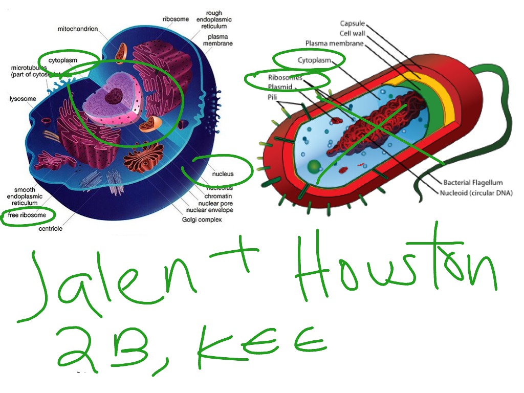
41 bacterial cell picture with labels
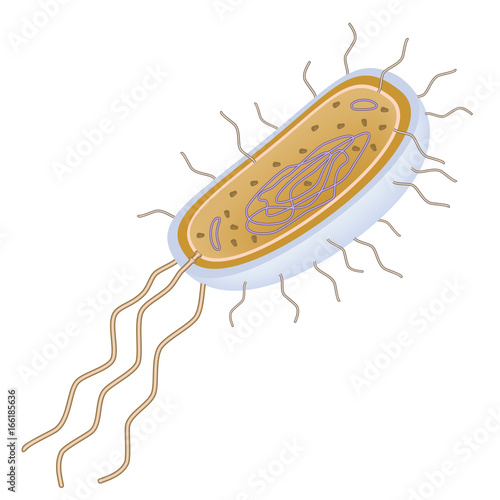
Label the Bacterium Cell - EnchantedLearning.com The cell is the basic unit of life. The following is a glossary of Bacterium cell terms. basal body - A structure that anchors the base of the flagellum and allows it to rotate. capsule - A layer on the outside of the cell wall. Most but not all bacteria have a capsule. cell wall - A thin membrane located outside the plasma membrane and within ... Bacterial Cell Structure Labeling Diagram | Quizlet Bacterial Microcompartment. Protein coated packets used to localize enzymes and other proteins into the cytoplasm. Plasmid. Double-stranded DNA circle containing extra genes. Flagella. specialized appendage attached to the cell by a basal body that holds a long rotating filament. Pushes cell forward. Endospore.
how to draw & label bacteria - YouTube | Science diagrams, Teaching, Labels Line Art. Hand Drawn. Royalty Free Stock Photos. How To Draw Hands. Medical. Cartoon. Find Set Different Bacteria Viruses Medical Info stock images in HD and millions of other royalty-free stock photos, illustrations and vectors in the Shutterstock collection. Thousands of new, high-quality pictures added every day. I.

Bacterial cell picture with labels
Plant and Animal Cells - Labeled Graphics A compilation of plant and animal cell images with organelles and major structures labeled. Students can print images to help them learn the cell. ... if students missed the lab that day they can view a site with pictures to complete lab handout Plant Cell ... looks at cheek and onion cells. Prokaryote Coloring - color a typical bacteria cell ... Structure of Bacteria (With Diagram) | Microbiology 8. According to Peberdy (1980) the only compound present in the cell walls of both Gram-negative and Gram-positive bacteria is 'peptidoglycan'. The cell walls of Gram-positive bacteria contain up to 95% peptidoglycan and up to 10% teichoic acids. 9. Cytoplasmic membrane is a thin (5-10 nm) layer lining the inner surface of the cell wall. A Labeled Diagram of the Animal Cell and its Organelles A Labeled Diagram of the Animal Cell and its Organelles. There are two types of cells - Prokaryotic and Eucaryotic. Eukaryotic cells are larger, more complex, and have evolved more recently than prokaryotes. Where, prokaryotes are just bacteria and archaea, eukaryotes are literally everything else. From amoebae to earthworms to mushrooms, grass ...
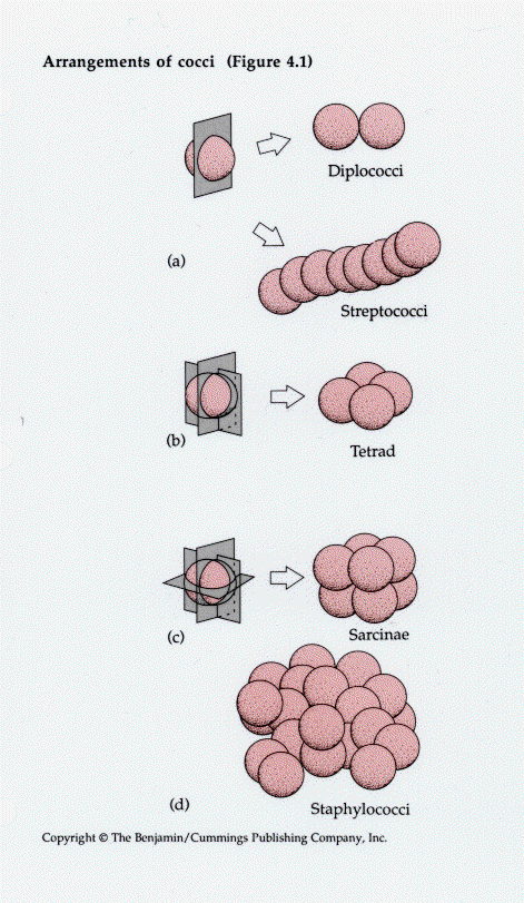
Bacterial cell picture with labels. Label bacteria cell - Teaching resources - Wordwall 4/2 Year 12 Prokaryotic Cell Label Quiz. by Brownc5. KS5 Y12. 6.1 Label the electrolysis cell Labelled diagram. by Kreeves2. Bacteria Hangman Hangman. by Shonprebble. KS3 Y8 Biology Science. Copy of Yr7 Animal Cell to Label Labelled diagram. Bacteria in Microbiology - shapes, structure and diagram Bacterial spores. Bacterial endospores layers. Bacteria cells are the smallest living cells that are known; even though viruses are smaller than bacteria, viruses are not living cells. There are different types of bacteria with various sizes, shapes, and structures. The bacteria shapes, structure, and labeled diagrams are discussed below. Structure of Bacterial Cell (With Diagram) - Biology Discussion Cell wall: It is a tough and rigid structure of peptidoglycan with accessory specific materials (e.g. LPS, teichoic acid etc.) surrounding the bacterium like a shell and lies external to the cytoplasmic membrane. It is 10-25 nm in thickness. It gives shape to the cell. Nucleus: The single circular double-stranded chromosome is the bacterial genome. Bacterial Cell Structure Diagram - Quizlet PLAY. Bacteria. Domain of unicellular prokaryotes that have cell walls containing peptidoglycan. Flagella. a slender whip-like structure that enables the bacteria to propel itself; an organ of locomotion. Ribosome. a cytoplasmic nuclear protein that is required in mRNA translation and protein synthesis. Nucleoid.
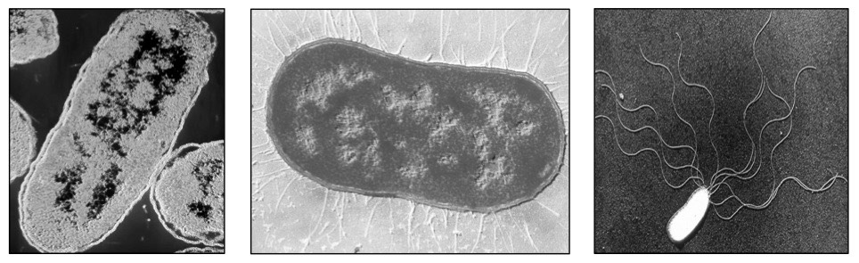
Image Library | CDC Online Newsroom | CDC Under a high magnification of 21674X, this digitally-colorized, scanning electron microscopic (SEM) image depicts a view of a dividing, Escherichia coli bacterium, clearly displaying the point at which the bacteria's cell wall was splitting into two separate organisms. See PHIL 7137 for a black and white version of this image. Bacterial Staining Microbiology Images ... - Science Prof Online 1. Endospore stain of Bacillus subtilis showing both endospores (green) & vegetative cells (pink) @1000xTM; 2. Negative endospore stain showing only vegetative cells @1000xTM; 3. Malachite green primary staining step of endopore stain with slide being heated over water bath; 4. Applying counterstain (safrinin) to bacterial smear as last step of ... 600+ Free Bacteria & Virus Images - Pixabay Bacteria and virus high resolution images. Find your perfect picture for your project. 639 Free images of Bacteria / 7 ‹ › ... A Labeled Diagram of the Animal Cell and its Organelles A Labeled Diagram of the Animal Cell and its Organelles. There are two types of cells - Prokaryotic and Eucaryotic. Eukaryotic cells are larger, more complex, and have evolved more recently than prokaryotes. Where, prokaryotes are just bacteria and archaea, eukaryotes are literally everything else. From amoebae to earthworms to mushrooms, grass ...
Structure of Bacteria (With Diagram) | Microbiology 8. According to Peberdy (1980) the only compound present in the cell walls of both Gram-negative and Gram-positive bacteria is 'peptidoglycan'. The cell walls of Gram-positive bacteria contain up to 95% peptidoglycan and up to 10% teichoic acids. 9. Cytoplasmic membrane is a thin (5-10 nm) layer lining the inner surface of the cell wall. Plant and Animal Cells - Labeled Graphics A compilation of plant and animal cell images with organelles and major structures labeled. Students can print images to help them learn the cell. ... if students missed the lab that day they can view a site with pictures to complete lab handout Plant Cell ... looks at cheek and onion cells. Prokaryote Coloring - color a typical bacteria cell ...
World Science Articles: Microbiology

A Connective Tissue Mast-Cell-Specific Receptor Detects Bacterial Quorum-Sensing Molecules and ...
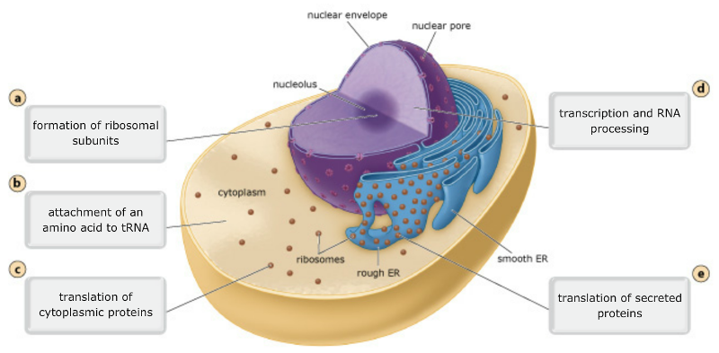
Print Exam 3: Ch. 17: From Gene to Protein flashcards | Easy Notecards

BACTERIAL CELL THING-LINK

Hyalomatrix®

Games: All

The Prokaryotic Cell (Bacteria) - PurposeGames
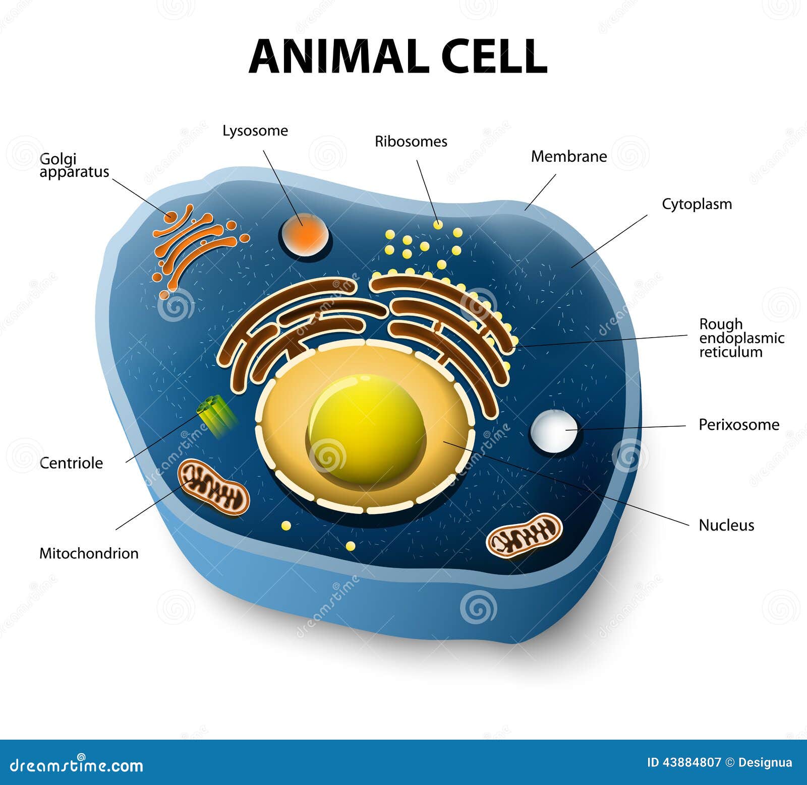
Animal Cell Cut-away Stock Vector - Image: 43884807

33 Label A Bacterial Cell - Labels For You
Microbiology Images: Science Photographs from Science Prof Online

Algae
Review

32 Label A Bacterial Cell - Labels Database 2020
ShowMe - Bacterial cells
Designua's "Microbes and Parasites (viruses and bacteria)" set on Shutterstock
Designua's "Microbes and Parasites (viruses and bacteria)" set on Shutterstock
Topic 1.2 Ultra-Structure of Cells - AMAZING WORLD OF SCIENCE WITH MR. GREEN
Bacterial Cell Diagram Unlabeled ~ DIAGRAM
Post a Comment for "41 bacterial cell picture with labels"